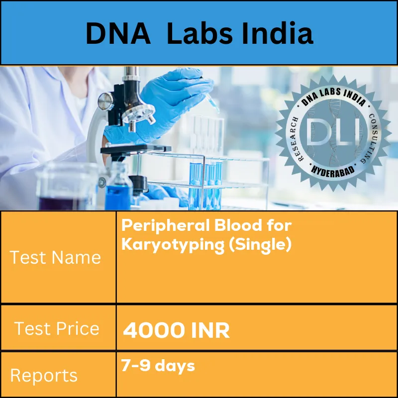
Peripheral blood for karyotyping (SINGLE)

Peripheral blood for karyotyping (SINGLE)

Disease: Genetics

Method: Cell culture

DNA Labs India is Ranked as No1 genetic DNA Test lab- 3500 Sample collection centers across India Call 07941057551 to talk with Doctor to get second opinion for free of cost - 100% All Conversation are private and confidential

Call 07941057551 Dr Vijaya

Sample Types
  • Blood
  • Blood Sodium Heparin

4,000.00/- Rs ₹5,000.0020% off

  • Results in : 7-9 days

Why to get tested at DNA Labs India for Peripheral blood for karyotyping (SINGLE) ?

Book Now
  • 3500 Sample collection centers, Free Home Sample collection for Peripheral blood for karyotyping (SINGLE) in your city
  • Ranked India No1 DNA Test Lab for Peripheral blood for karyotyping (SINGLE)
  • Most Trusted Lab by doctors for Peripheral blood for karyotyping (SINGLE). Call 07941057551 to talk with Doctor to get second opinion for free of cost
  • 100% All Conversation are private and confidential Call 07941057551 to talk with doctor 100% Accurate Test Results for Peripheral blood for karyotyping (SINGLE)

Peripheral blood for karyotyping (SINGLE) Cost 4000 Rs


Test Name Peripheral blood for karyotyping (SINGLE)
Test type Gynecologist
Pre-test Information Peripheral blood for karyotyping (SINGLE) is performed using Cell culture methodology. Sample that have to be given is Peripheral blood in Sodium Heparin Vacutainer (2ml) for this test. You can expect results in 7-9 days
Report Delivery 7-9 days
Components Sodium Heparin Vacutainer (2ml)
Price ₹ 4000
Method Cell culture

Peripheral blood for karyotyping (SINGLE) Details


Short Description

Doctor recommend karyotyping test to detect chromosome abnormalities, thus to help diagnose genetic diseases, some congenital disabilities, and certain disorders of the blood and lymphatic system When pregnancy screening tests are abnormal Whenever signs of a chromosomal abnormality-associated disorder are present Bad obstetric history Recurrent pregnancy loss Infertility Primary amenorrhea Dysmorphic features of a newborn When signs of a chromosomal abnormality or associated disorder are present When a specific abnormality has been detected in a family member When a person has leukaemia, lymphoma, myeloma, myelodysplasia or another cancer and an acquired chromosome abnormality is suspected

Test Specifications

  • Speciality: Gynecologist

  • Components: Sodium Heparin Vacutainer (2ml)

  • Department:

  • Shipping Stability: Ambient

PreTest Information

Peripheral blood for karyotyping (SINGLE) is performed using Cell culture methodology. Sample that have to be given is Peripheral blood in Sodium Heparin Vacutainer (2ml) for this test. You can expect results in 7-9 days

Detail Description

Karyotyping chromosome analysis Test Details:

What type of sample is required for karyotyping?

  • A blood sample is drawn from a vein in your arm;
  • a sample of amniotic fluid or chorionic villus from a pregnant woman;
  • a bone marrow or tissue sample

What is being tested in Karyotyping?

In Karyotyping lymphocytes from peripheral blood are cultured, using mitogens to stimulate the transformation of the lymphocytes into mitotically active cells. The timing of harvesting of the cells is engineered such that a maximum number of cells are in metaphase.

The cells are then fixed and spread onto a slide. The chromosomes are stained using many stains, usually, Giemsa  (G-banding and R-banding), which produces banding patterns on the chromosomes with a band resolution of 400 – 650 bands per haploid chromosome set.

The chromosomes with their bands are then examined microscopically for abnormalities such as loss or gain of entire chromosomes, translocations of all or part of an arm of one chromosome to another, or more subtle changes in banding patterns associated with various genetic syndromes. The chromosomes are photographed and rearranged into pairs for examination (a karyogram).

The benefits of karyotyping are:

  1. It can view the entire genome.
  2. It can visualize individual cells and individual chromosomes.

The limits of karyotyping are:

  1. Resolution limited to around 5 Mb.
  2. An actively growing source of cells is required.

It is important to note that classic karyotyping is timeconsuming, with the preparation of cells for examination taking several days. Also, live lymphocytes are required, so blood samples need to arrive at the laboratory within a maximum of 48 hours after sampling, preferably sooner, to avoid failure of cell growth in culture.

The test is performed by:

  • Taking a sample of a person’s cells, culturing them in nutrient-enriched media to promote cell division in vitro. This is done to select a specific time during the cells’ growth phase when the chromosomes are easiest to distinguish.
  • Isolating the chromosomes from the nucleus of the cells, placing them on a slide, and treating them with a particular stain.
  • Taking microphotographs of the chromosomes.
  • In jigsaw puzzle fashion, rearranging the pictures of the chromosomes to match up pairs and arrange them by size, from largest to smallest, numbers 1 to 22, followed by the sex chromosomes as the 23rd pair.
  • The pictures also allow the chromosomes to be vertically oriented. Each chromosome looks like a striped straw. It has two arms that differ in length (a short arm (p) and a long arm (q)), a pinched-in area between the components called a centromere, and a series of light and dark horizontal bands. The length of the arms and the location of the bands help determine top from bottom.
  • Once the chromosome photo arrangement is completed, a laboratory specialist evaluates the chromosome pairs and identifies any abnormalities that may be present.

Some chromosomal disorders that may be detected include:

  • Down syndrome (Trisomy 21), caused by an extra chromosome 21; this may occur in all or most cells of the body.
  • Edwards syndrome (Trisomy 18), a condition associated with severe mental retardation; caused by an extra chromosome 18.
  • Patau syndrome (Trisomy 13), caused by an extra chromosome 13.
  • Klinefelter syndrome, the most common sex chromosome abnormality in males; caused by an extra X chromosome.
  • Turner syndrome, caused by missing one X chromosome in females.
  • Chronic myelogenous leukaemia, a classic 9;22 translocation that is diagnostic of the disease.

How is the sample collected for testing?

  • A blood sample is obtained by inserting a needle into a vein in the arm.
  • Amniotic fluid and chorionic villi are collected from a pregnant woman by a healthcare practitioner using amniocentesis or chorionic villus sampling procedures.
  • Bone marrow or tissue sample collections require a biopsy procedure to be performed.


How is the karyotyping test used?

A chromosomal karyotype is used to detect chromosome abnormalities and thus used to diagnose genetic diseases, some congenital disabilities, and certain disorders of the blood or lymphatic system.

It may be performed for:

  • A fetus, using amniotic fluid or chorionic villi (tissue from the placenta):
    • If one or more of a woman’s pregnancy screening tests, such as the first trimester Down syndrome screen or the second-trimester maternal serum screening, are abnormal.
    • If a pregnant woman is having amniotic fluid analysis performed because she is considered at higher than usual risk of having a baby with a congenital disability.
    • If fetal structural and developmental abnormalities are detected, such as during an ultrasound.
    • If there is a known chromosomal abnormality in the family line.
  • A woman or a couple, before pregnancy, to evaluate her or their chromosomes, especially if a woman has experienced previous miscarriages or infertility.
  • Tissue from a miscarriage or stillbirth, to help determine if the cause was due to a chromosomal abnormality in the fetus.
  • An infant who is born with congenital abnormalities, including congenital physical disabilities, mental retardation, delayed growth and development, or signs of a specific genetic disorder.
  • A person with infertility or one who shows signs of a genetic disorder.
  • Family members, to detect specific chromosomal abnormalities when they have been seen in a child or another family member.
  • A person who has been diagnosed with certain types of leukaemia, lymphoma, refractory anaemia, or cancer as these conditions can lead to acquired changes in chromosomes; this testing may be performed on blood or a bone marrow sample.

When is karyotyping test ordered?

Chromosome analysis may be ordered when a fetus is suspected of having a chromosomal abnormality when an infant has congenital abnormalities, when a woman experiences miscarriages or infertility, and when an adult shows signs of a genetic disorder.

It may also be ordered to detect the presence of a chromosomal abnormality in family members when it has been seen in a child or another family member.

It may be ordered to detect acquired chromosomal abnormalities when an individual has leukaemia, lymphoma, myeloma, refractory anaemia, or another cancer.

What does the karyotyping test result mean?

Interpretation of test results must be made by a person with specialized training in cytogenetics. Some findings are relatively straightforward, such as an extra chromosome 21 (Trisomy 21) indicating Down syndrome, but others may be very complex.

Although there will be typical signs with specific chromosomal abnormalities, the effects and the severity may vary from person to person and often cannot be reliably predicted.

Some examples of abnormalities that chromosome analysis may reveal include:

Trisomy This is the presence of an extra chromosome, a third instead of a pair. Diseases associated with trisomies include Down syndrome (related to a Trisomy of chromosome 21), Patau syndrome (Trisomy 13), Edward syndrome (Trisomy 18), and Klinefelter syndrome (a male with an extra X chromosome – XXY instead of XY).
Monosomy This is the absence of one of the chromosomes. An example of monosomy is Turner syndrome (a female with a single X chromosome – X instead of XX). Most other monosomies are not compatible with life.
Deletions These are missing pieces of chromosomes and genetic material. Some may be small and difficult to be detected.
Duplications These represent extra genetic material and may be present on any chromosome, such as the presence of two horizontal bands at a specific location instead of one.
Translocations With translocations, pieces of chromosomes break off and reattach to another chromosome. If it is a one-to-one switch and all of the genetic material is present (but in the wrong place), it is said to be a balanced translocation. If it is not, then it is called an unbalanced translocation.
Genetic Rearrangement With this, genetic material is present on a chromosome but not in its usual location. A person could have both rearrangement and duplication or deletion. An almost infinite number of rearrangements are possible. Interpreting the effects of the changes can be challenging.

Duplications, deletions, translocations, and genetic rearrangements can cause a myriad of health and development issues. It depends upon what genes are missing or are present in too many copies.

Some genetic rearrangements will be variations that do not cause noticeable symptoms. Balanced translocations (where two chromosomes have swapped portions of themselves but all of the genetic material is present) may cause no problems for the person who has them but may cause problems in their children.

Many hematologic and lymphoid malignancies (e.g., leukaemia, lymphoma, myeloma, myelodysplasia) are associated with chromosomal abnormalities, which can help diagnose the disease and predict the clinical course of the disease.

Why do the drawbacks of karyotyping test?

Some chromosome alterations are too small or subtle to detect with karyotyping. Other testing techniques such as fluorescent in situ hybridization (FISH) or a microarray may sometimes be performed to investigate chromosomal abnormalities further.

People can have cells in their body with different genetic material. This happens because of changes early in the development of a fetus that leads to the development of distinctly different cell lines and is called mosaicism. An example of this is some cases of Down syndrome. The affected person can have some cells with an extra third chromosome 21 and some cells with the standard pair.

Should everyone have a karyotyping test done?

Chromosome analysis is frequently performed, but it is not indicated as a general screening test. The majority of people will never need to have one done.

Why does the karyotype take several days to perform?

The cells that are tested must be cultured and cell division promoted. The amount of time that this takes will vary from sample to sample. Highly sophisticated, abnormal karyotypes may require a longer time to evaluate.

Frequently Asked Questions

  • What is the cost of Peripheral blood for karyotyping (SINGLE)?

    Cost of Peripheral blood for karyotyping (SINGLE) is 4000 Rs

₹4,000.00 ₹5,000.0020% off

Book Now
  • Free Home Sample collection
  • Ranked India No1 DNA Test Lab
  • Most Trusted Lab by doctors
  • 100% Accurate Test Results