Understanding Macular Atrophy: Beyond the Basics of Vision Loss
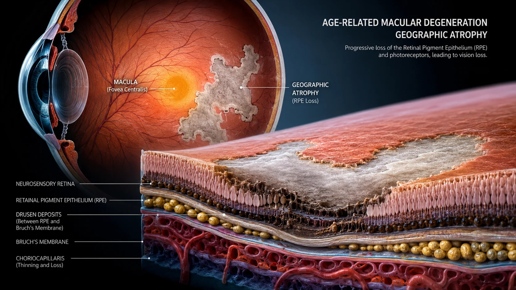
Macular atrophy represents a significant challenge in eye health, characterized by the progressive and irreversible loss of cells in the macula—the central part of the retina responsible for sharp, detailed vision. As an advanced form of age-related macular degeneration (AMD), it leads to permanent impairment of activities like reading, driving, and recognizing faces .
This condition affects approximately 5 million people globally, with prevalence increasing exponentially with age . The impact is profound, as 67% of patients with geographic atrophy become ineligible to drive within a median of 1.6 years based on visual acuity alone .
The Biology of Macular Atrophy: What Actually Happens in Your Eye
To understand macular atrophy, it helps to first appreciate the macula’s role. The macula is the central region of the retina—the light-sensitive tissue at the back of the eye. It enables us to see fine details, read, and recognize faces.
Macular atrophy occurs when the retinal pigment epithelium (RPE)—a supportive layer beneath the photoreceptors—deteriorates and dies. This leads to the progressive loss of photoreceptors (the cells that detect light) and the choriocapillaris (the underlying blood vessel network) in well-defined patches within the macula .
The term “geographic atrophy” is often used interchangeably with macular atrophy, referring to the late stage of dry AMD . The “geographic” descriptor comes from the appearance of these atrophic areas as sharply defined, map-like lesions that expand over time .
Drusen: The Warning Signs
The onset of macular atrophy is typically preceded by the accumulation of drusen—lipid-rich extracellular deposits that form beneath the retina. These yellow deposits can be detected during routine eye examinations and are classified by size:
- Grade 1 (Early AMD): Few drusen deposits smaller than 63 µm
- Grade 2-3 (Intermediate AMD): Drusen between 63-124 µm
- Grade 4 (Geographic Atrophy): Drusen >125 µm with distinct areas of RPE and photoreceptor loss
Two Distinct Pathways to Atrophy: New Research Insights
Recent clinical research has revealed that macular atrophy can develop through two separate pathways, even in patients receiving treatment .
Pathway 1: Neovascular-Related Atrophy
This pathway is associated with the development and exudation of macular neovascularization (MNV)—abnormal blood vessel growth commonly seen in wet AMD. Causes include:
- Physical harm from Type 3 neovessels
- Collapse of serous pigment epithelium detachment
- Fibrosis (scar tissue formation)
Pathway 2: Non-Neovascular Atrophy
This pathway involves lesions unrelated to the neovascularization process itself, primarily:
- Drusen accumulation
- Subretinal drusenoid deposits (reticular pseudodrusen)
In a study of 41 eyes with Type 3 MNV, macular atrophy was present in 92.7% after 12 months of anti-VEGF therapy, with precursors split between these two pathways .
Global and Indian Perspectives: Who Is Affected?
Macular atrophy affects diverse populations differently, with important implications for Indian patients.
Prevalence Among Asian Populations
A pooled analysis from the Asian Eye Epidemiology Consortium, including data from rural India, found the prevalence of myopic macular degeneration (MMD)—which includes macular atrophy—to be 0.4% among Asians in rural India . This compares to:
- 0.5% in Beijing, China
- 1.5% in Russia
- 5.2% in Singapore
The Central India Eye and Medical Study (2006), involving 4,561 participants over age 30, reported a macular atrophy prevalence of 0.2% .
Key Risk Factors for Asian Populations
The Asian Eye Epidemiology Consortium identified several significant predictors for macular atrophy:
- Older age (per year; OR=1.13)
- Female sex (OR=2.0)
- Myopia severity (per negative diopter; OR=1.7)
- Longer axial length (per mm; OR=3.1)
- Lower education (OR=1.9)
Recognizing the Signs: Symptoms of Macular Atrophy
Macular atrophy typically develops gradually, with symptoms becoming more pronounced as the fovea (the central point of the macula) becomes involved .
Common Symptoms
- Loss of central vision acuity: Difficulty reading, recognizing faces, or performing detailed tasks
- Dark spots or scotomas: Blind spots in the central field of vision
- Difficulty in dim light: Needing brighter illumination for everyday activities
- Duller color perception: Colors appearing less vibrant or faded
The Silent Progression
Early macular atrophy may be asymptomatic, particularly if it begins in one eye or spares the fovea (a phenomenon known as “foveal sparing”). The atrophic patches typically appear in the perifoveal macula initially and expand over 1.5 to 2.4 years to involve the fovea .
How Is Macular Atrophy Diagnosed?
Early detection is critical for managing macular atrophy and preserving vision. Diagnosis involves a comprehensive eye examination and advanced imaging techniques .
Key Diagnostic Tools
Fundus Autofluorescence (FAF): A non-invasive imaging technique that detects lipofuscin (a fluorescent pigment) accumulation in the RPE. Areas of atrophy appear as regions with reduced autofluorescence signals due to RPE cell loss .
Optical Coherence Tomography (OCT): Provides high-resolution cross-sectional images of the retina, allowing clinicians to visualize retinal layers and detect early structural changes, including photoreceptor loss and RPE atrophy .
Color Fundus Photography: Captures two-dimensional images of the retina to visualize drusen, pigment changes, and atrophy .
Microperimetry: Assesses macular sensitivity by projecting light stimuli onto different retinal points, generating a functional “map” of vision .
The Importance of Comprehensive Assessment
While visual acuity testing is important, it does not fully capture the patient’s visual experience. A comprehensive assessment should combine:
- Best corrected visual acuity (BCVA)
- Low-luminance visual acuity (LLVA)
- Microperimetry
- Reading speed tests
Treatment and Management Strategies
Recent years have brought significant advances in macular atrophy treatment, though a cure remains elusive.
FDA-Approved Treatments
Two complement inhibitor therapies have been approved to slow disease progression:
Pegcetacoplan (SYFOVRE™) and avacincaptad pegol (IZERVAY™) are administered as intraocular injections monthly or every other month. These medications target the complement cascade—a part of the immune system implicated in the inflammatory processes driving AMD progression .
While these treatments show promising results in reducing lesion growth, they do not reverse existing damage and carry safety considerations .
Supportive Therapies
- AREDS2 Supplements: A specific combination of antioxidants (lutein, zeaxanthin, vitamin C, vitamin E, zinc, and copper) may slow progression in some patients .
- Visual Rehabilitation: Low-vision aids, special eyewear, and magnifying glasses can help maintain independence .
- Implantable Miniature Telescope (IMT): A surgical option that magnifies central vision and focuses it on healthy areas of the retina .
Clinical Trials and Emerging Approaches
Numerous clinical trials are exploring new therapeutic strategies, including:
- Anti-inflammatory therapies
- Neuroprotective agents
- Lipofuscin and visual cycle inhibitors
- Choroidal blood flow restoration
- Gene therapy
Lifestyle Modifications: Reducing Your Risk
While macular atrophy cannot be entirely prevented, addressing modifiable risk factors can reduce your likelihood of developing the condition .
Key Lifestyle Recommendations
- Quit smoking: Smoking is a major modifiable risk factor for AMD and macular atrophy
- Manage chronic conditions: Control diabetes, high blood pressure, high cholesterol, and obesity
- Protect your eyes: Wear sunglasses with yellow tint for sun protection; use safety eyewear for work and sports
- Eat a healthy diet: Consume dark green leafy vegetables and foods rich in antioxidants
- Regular eye exams: Annual comprehensive eye examinations are essential for those over 50
When to See an Eye Specialist
Prompt evaluation is crucial if you experience:
- Blurred or distorted central vision
- Difficulty reading, recognizing faces, or driving
- Dark spots or blind spots in your central vision
- Colors appearing dull or faded
- Increased difficulty seeing in low light
These symptoms could indicate macular atrophy or other serious eye conditions requiring immediate attention .
Disclaimer: This content is for informational purposes only and does not constitute medical advice. Always consult with a qualified healthcare provider regarding diagnosis and treatment options for any medical condition.